微创血管内介入栓塞术治疗颅脑动脉瘤临床研究
黄春波1) 杨瑞生1) 黄亚楠2) 李庆安1) 王伟丰1) 靳晓亮1)
济源市人民医院 1)神经外科 2)骨科,河南 济源 459000
作者简介:黄春波,Email:15978718836@163.com
【摘要】 目的 探讨微创血管内介入栓塞术对颅脑动脉瘤患者术后脑组织血流量及生存质量的影响。方法 选取济源市人民医院81例颅脑动脉瘤患者,根据手术术式不同分为对照组40例,观察组41例,对照组予以开颅夹闭术治疗,观察组予以微创血管内介入栓塞术治疗,观察2组手术前后脑组织血流量(CBF)变化情况、并发症发生情况及手术前后生存质量评分。结果 阻断颅内动脉瘤时及术后观察组CBF均低于对照组,且2组阻断颅内动脉瘤时、术后CBF均较术前有所变化,但观察组变化幅度小于对照组,差异具有统计学意义(P<0.05);观察组并发症发生率为4.88%(2/41),低于对照组22.50%(9/40),差异具有统计学意义(P<0.05);术后观察组生存质量各指标评分均高于对照组,差异具有统计学意义(P<0.05)。结论 微创血管内介入栓塞术对颅脑动脉瘤患者术后脑组织血流量影响小,可提高患者生存质量,降低并发症发生率。
【关键词】 微创血管内介入栓塞术;颅脑动脉瘤;脑组织血流量;生存质量;并发症
【中图分类号】 R739.4 【文献标识码】 A 【文章编号】 1673-5110(2018)18-2054-09 DOI:10.12083/SYSJ.2018.18.450
Clinical study on minimally invasive endovascular interventional embolization in the treatment of craniocerebral aneurysms
HUANG Chunbo1),YANG Ruisheng1),HUANG Yanan2),LI Qing’an1),WANG Weifeng1),JIN Xiaoliang1)
1)Department of Neurosurgery,the People's Hospital of Jiyuan,Jiyuan 459000,China;2)Department of Orthopedics,the People's Hospital of Jiyuan,Jiyuan 459000,China
【Abstract】 Objective To investigate the effect of minimally invasive endovascular interventional embolization on postoperative cerebral blood flow and quality of life in patients with craniocerebral aneurysms.Methods A total of 81 cases of craniocerebral aneurysm in our hospital were divided into the control group of 40 cases and the observation group of 41 cases.The control group was treated with craniotomy,and the observation group was treated with minimally invasive endovascular interventional embolization.The changes of cerebral blood flow (CBF) before and after operation,the occurrence of complications and the quality of life before and after operation were compared between the two groups.Results At the time when the intracranial aneurysms were blocked and after operation,the CBF in the observation group were lower than that in the control group.And at the time when the intracranial aneurysms were blocked and after operation,the CBF in two group changed compared with that before operation,but the change range of the observation group was smaller than that of the control group,and the difference was statistically significant(P<0.05).The incidence of complications in the observation group was 4.88% (2/41),which was lower than that of the control group (22.50% (9/40)),and the difference was statistically significant(P<0.05).The score of the quality of life of the observation group was higher than that of the control group after the operation,and the difference was statistically significant(P<0.05).Conclusion Minimally invasive endovascular interventional embolization has little effect on cerebral blood flow in patients with craniocerebral aneurysms after operation.It can improve the patient's quality of life and reduce the incidence of complications.
【Key words】 Minimally invasive endovascular interventional embolization;Craniocerebral aneurysm;Cerebral blood flow;Quality of life;Complication
颅脑动脉瘤为临床常见脑部出血性疾病,相关数据显示,在非外伤性蛛网膜下腔出血中高达80%为动脉瘤破裂引起[1-2]。颅脑动脉瘤具有恶性程度高、致残致死率高、病情进展快等特点,故患者需及时实施手术治疗[3]。开颅夹闭瘤颈术为目前临床治疗颅脑动脉瘤常用手术术式,可有效降低病死率,提高患者生活质量,但该术式对患者造成创伤大,手术风险性大,且易损害周围正常脑组织,术后恢复慢,预后效果较差[4-6]。有学者指出,微创血管内介入栓塞术治疗颅脑动脉瘤,效果较佳,不仅可降低致残致死率,且可使绝对危险度降低7.4%左右,利于术后恢复[7]。本研究选取81例颅脑动脉瘤患者,分为2组,并予以不同手术术式治疗,观察微创血管内介入栓塞术对颅脑动脉瘤患者术后脑组织血流量及生存质量的影响。
1 资料和方法
1.1 一般资料 选取济源市人民医院2013-10—2016-09收治的81例颅脑动脉瘤患者,根据手术术式不同分为对照组40例,男25例,女15例,年龄35~65(53.58±5.22)岁,Hunt-Hess分级[8]:Ⅰ级15例,Ⅱ级13例,Ⅲ级12例;观察组41例,男24例,女17例,年龄36~63(54.61±5.19)岁,Hunt-Hess分级:Ⅰ级16例,Ⅱ级14例,Ⅲ级11例。对比2组年龄、性别、Hunt-Hess分级等基线资料,差异无统计学意义(P>0.05)。
1.2 纳入及排除标准 (1)纳入标准:经数字减影血管造影或颅脑CT诊断并确诊为颅脑动脉瘤;年龄35~70岁;知晓本研究并签署知情同意书。(2)排除标准:合并肝、肾等多脏器功能衰竭者;合并传染性疾病者;复发性动脉瘤;同时实施两种以上方法治疗者;哺乳期及妊娠期女性;存在严重意识障碍或其他因素导致难以完成本研究者。
1.3 手术方法 均予以降低颅内压、抗脑血管痉挛、防止再出血、脑脊液引流、维持酸碱及水电解质平衡等常规治疗,呼吸困难者予以呼吸机辅助呼吸。对照组实施开颅夹闭手术治疗:行全麻,气管插管,对颅内动脉瘤进行定位,自翼点入路,于头部行7~8 cm弧形切口,依次切开皮肤、皮下组织、骨膜,将皮瓣翻转,于颞上线与冠状缝交界处对颅骨进行钻孔,以铣刀铣下骨窗,将硬膜剪开,顺脑裂进行分离直至动脉瘤,采用血管夹阻断载瘤动脉,对动脉瘤及其周围粘连进行分离,选取适宜动脉瘤夹夹闭动脉瘤,将阻断夹去除,观察瘤颈夹闭情况,硬膜下放置引流管,逐层缝合切口,术后2 d拔除引流管。观察组实施微创血管内介入栓塞术:术前严密监测心率、血压等生命体征,同时进行全身肝素化2 h,术中给予1.25 g/h肝素钠,全麻,于右侧腹股沟处股动脉实施seldenger技术穿刺,置入6F动脉鞘,并将导引导管放入同侧股动脉,导管头端放置于颈内动脉内,微导管于微导丝引导下插入,微导管头端于数字减影血管造影路径图引导下置入动脉瘤内中、外约1/3处,选取与动脉瘤大小相当的弹簧圈填塞,松弛导管确定稳定后解脱。采用Onyx胶栓塞者,于微导管置入瘤体后根据造影结果确认瘤体及其远端无血管分支,以氯化钠溶液(0.9%)冲洗导管,采用注射器将二甲基亚砜注射入导管,吸取Onyx-18胶,并注入瘤体腔内,有少许反流出现则停止推注;同时监测数字减影血管造影,若为夹层动脉瘤或宽颈或动脉瘤较大则应实施Rimording技术栓塞或采用支架辅助栓塞,直至动脉瘤不显影后拔出导管,术后使用沙袋压迫穿刺部位,术后12 h皮下注射5 000 U低分子肝素。术后均严密监测生命体征、抗血管痉挛、控制血压及抗生素抗感染等治疗。
1.4 观察指标 (1)观察比较2组术前、阻断颅内动脉瘤时及术后患者脑组织血流量(CBF)变化情况。(2)观察比较2组肺部感染、脑积水、血管痉挛、尿路感染等并发症发生情况。(3)采用生存质量评分量表(SF-36)对2组手术前后社会功能、生理功能、活力、健康状况等8个方面生存质量进行评估比较,得分越高,生存质量越好[9]。
1.5 统计学分析 运用SPSS 19.0软件进行数据分析,计量资料以均数±标准差(x±s)表示,采用t检验,计数资料以率(%)表示,采用卡方检验,P<0.05为差异有统计学意义。
2 结果
2.1 2组手术前后CBF变化情况比较 术前2组CBF比较,差异无统计学意义(P>0.05),阻断颅内动脉瘤时及术后观察组CBF均低于对照组,且2组阻断颅内动脉瘤时、术后CBF均较术前有所变化,但观察组变化幅度小于对照组,差异具有统计学意义(P<0.05)。见表1。
表1 2组手术前后CBF变化情况对比 [x±s,mL/(100 g·min)]
Table 1 Comparison of CBF changes before and after operation in 2 groups [x±s,mL/(100 g·min)]
| 组别 |
n |
术前 |
阻断颅内动脉瘤时 |
术后 |
| 观察组 |
41 |
55.59±8.82 |
58.71±9.40 |
56.19±8.80 |
| 对照组 |
40 |
56.91±8.90 |
69.41±8.39 |
60.48±8.51 |
| t值 |
|
0.67 |
5.4 |
2.23 |
| P值 |
|
>0.05 |
<0.05 |
<0.05 |
2.2 2组术后并发症比较 观察组并发症发生率低于对照组,差异具有统计学意义(P<0.05)。见表2。
2.3 2组手术前后生存质量评分比较 术前2组生存质量各指标评分对比,差异无统计学意义(P>0.05),术后观察组生存质量各指标评分均高于对照组,差异具有统计学意义(P<0.05)。见表3。
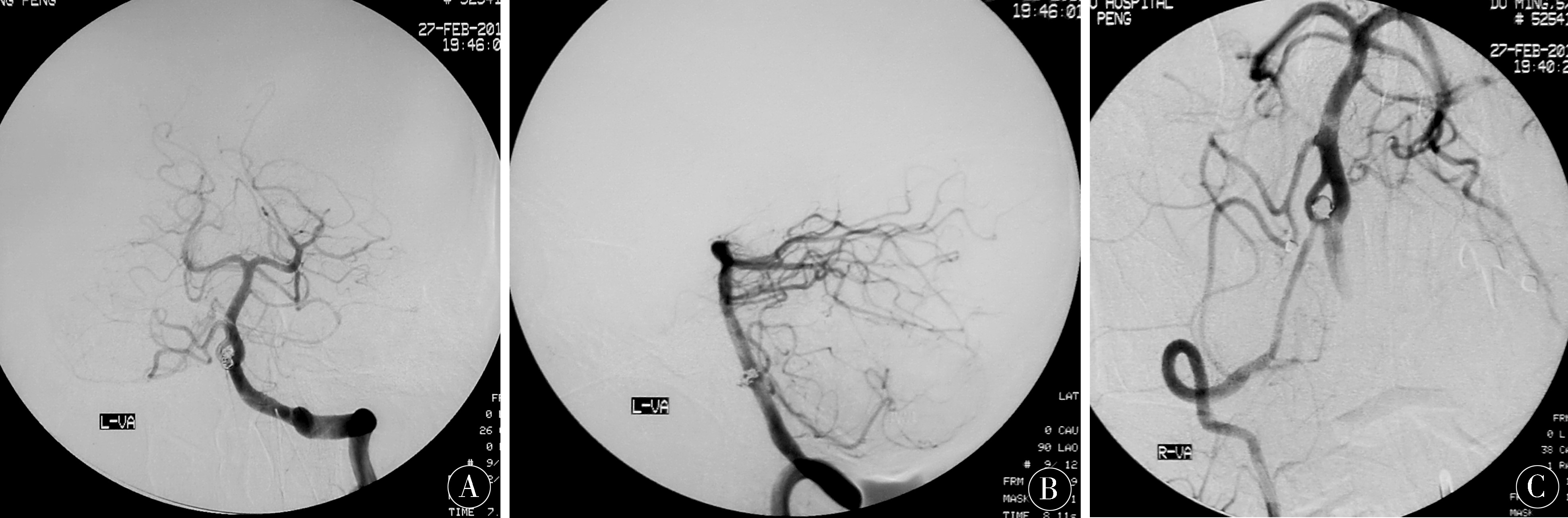
2.4 典型病例分析 动脉瘤完全栓塞,瘤颈无残留,且开窗的两支血管未见狭窄或闭塞。见图1。
表2 对比2组术后并发症发生率 [n(%)]
Table 2 Comparison of the incidence of postoperative complications in the 2 groups [n(%)]
| 组别 |
n |
肺部感染 |
脑积水 |
血管痉挛 |
尿路感染 |
并发症发生率 |
| 观察组 |
41 |
1(2.44) |
0 |
1(2.44) |
0 |
2(4.88) |
| 对照组 |
40 |
2(5.00) |
2(5.00) |
3(7.50) |
2(5.00) |
9(22.50) |
| χ2值 |
|
|
|
|
5.357 |
|
| P值 |
|
|
|
|
<0.05 |
|
表3 2组手术前后生存质量评分 (x±s,分)
Table 3 Quality of life scores before and after surgery in 2 groups (x±s,score)
| 时间 |
组别 |
n |
社会功能 |
生理功能 |
健康状况 |
活力 |
生理职能 |
精神状况 |
情感功能 |
机体疼痛 |
| 术前 |
观察组 |
41 |
52.73±7.24 |
52.48±6.13 |
49.21±5.85 |
56.31±4.28 |
51.51±6.47 |
59.24±5.53 |
56.27±4.62 |
60.41±5.29 |
| |
对照组 |
40 |
52.50±7.16 |
51.27±7.50 |
50.23±4.75 |
55.13±4.24 |
51.46±6.21 |
59.33±5.48 |
55.20±4.71 |
60.67±5.18 |
| |
t值 |
|
0.144 |
0.796 |
0.86 |
1.246 |
0.036 |
0.074 |
1.032 |
0.223 |
| |
P值 |
|
>0.05 |
>0.05 |
>0.05 |
>0.05 |
>0.05 |
>0.05 |
>0.05 |
>0.05 |
| 术后 |
观察组 |
41 |
65.15±8.05 |
66.24±6.56 |
70.77±5.51 |
67.23±4.61 |
64.36±7.42 |
75.23±6.57 |
69.59±4.45 |
73.42±4.11 |
| |
对照组 |
40 |
58.36±8.39 |
56.29±7.31 |
60.76±5.23 |
57.69±4.24 |
54.24±7.21 |
64.63±6.09 |
60.69±5.65 |
67.93±4.10 |
| |
t值 |
|
3.717 |
6.451 |
8.382 |
9.678 |
6.223 |
7.526 |
7.886 |
6.018 |
| |
P值 |
|
<0.05 |
<0.05 |
<0.05 |
<0.05 |
<0.05 |
<0.05 |
<0.05 |
<0.05 |
图1 A、B:造影复查左侧椎动脉正侧位像图;C:右侧椎动脉正位像
Figure 1 A,B:Contrast examination of the left lateral vertebral artery positive lateral
image;C:Right vertebral artery orthotopic image
3 讨论
颅脑动脉瘤为临床常见脑血管疾病之一,被称为颅内的“不定时炸弹”,多数学者认为高血压、流体动力学效应、颅内动脉先天缺陷、动脉粥样硬化及胶原蛋白基因突变等为诱发颅内动脉瘤的主要因素[10-26];临床病理显示颅内血管同外周血管结构上的差异性、内外膜弹力组织相对较少、中层较为薄弱、动脉管壁退化及炎症反应、创伤引起的血管壁损伤等均可促进动脉瘤形成,此外脑组织血流量等血流动力学改变亦可诱发颅脑动脉瘤[27-40]。故选取一种合理、有效手术术式,改善患者脑血流动力学,控制疾病进展,降低并发症发生率,对患者具有重要意义。
开颅夹闭术具有术野清晰、手术成功率高等优势,为多发性动脉瘤、载瘤动脉解剖结构位置复杂、颅内形成血肿及瘤体较大患者首选手术术式,但该方法需于患者头颅部行7~8 cm长切口,创伤大,体质较弱者或老年患者难以耐受,且术后并发症发生率较高,延长患者住院时间,加重其经济负担[41-60]。张青松等[21]研究证实,血管内介入栓塞术治疗颅脑动脉瘤效果较佳,可有效改善预后,缩短治疗时间及住院时间,术后感染发生率仅为2.5%。微创血管内介入栓塞术是一种新兴微创手术术式,其通过穿刺股动脉,利用微导管及导管将弹簧圈置入动脉瘤内,充满动脉瘤,将动脉瘤囊内正常血运阻断,改善患者临床症状,避免发生动脉瘤破裂出血等不良事件,有效弥补了传统开颅手术术后并发症多的难题,且该术式无需开颅便可实施手术操作,对患者造成创伤小,对周围正常脑组织影响小,可减轻患者术中及术后疼痛程度,减少并发症发生率,促进良好预后[61-78];同时该术式适用范围广,适用于难以实施开颅手术治疗的宽颈或梭形动脉瘤及75岁以上老年患者;此外颅内动脉瘤患者入院时病情较为危重,全身情况差,可通过实施动脉瘤血管内栓塞,避免或减少出血的发生,为患者争取最佳治疗时机,降低手术风险,提高手术成功率[79-89]。本研究显示,术后观察组CBF均低于对照组,术前、阻断颅内动脉瘤时及术后2组CBF均有所改变,但观察组改变幅度小于对照组,且观察组并发症发生率低于对照组,提示对颅脑动脉瘤患者给予微创血管内介入栓塞术治疗,可有效降低并发症发生率,维持脑组织血流量稳定;同时本研究发现术后观察组生存质量各指标评分均高于对照组,旨在说明微创血管内介入栓塞术可改善颅脑动脉瘤患者生存质量。此外采用微创血管内介入栓塞术治疗时应及时释放颅内残存脑脊液,减少脑积水的发生;术前应对动脉瘤患者进行严格检查,对存在占位效应的动脉瘤应考虑采取开颅手术治疗,避免加重占位症状[90-105]。
微创血管内介入栓塞术应用于颅脑动脉瘤患者,可降低并发症发生率,提高患者术后生存质量,对脑组织血流量影响小,具有较高应用价值。
4 参考文献
[1] 荣向霞,黄冠敏,黄艳,等.血管内介入栓塞术治疗颅内动脉瘤的护理[J].安徽医学,2013,34(9):1 408-1 410.
[2] LIKA M,VULEV I,LISKOV Z,et al.Traumatic aneurysms of middle cerebral artery after a penetrating craniocerebral trauma[J].Ceska Radiologie,2017,71(1):41-46.
[3] 石会,赵兵,钟鸣,等.三维数字减影血管造影对颅内动脉瘤夹闭术后残留的评价及应用[J].实用医学杂志,2014,30(7):1 024-1 027.
[4] 王小霞,曹作为,夏鹰,等.颅内动脉瘤破裂经血管内介入栓塞治疗的临床护理[J].海南医学,2014,25(3):465-466.
[5] 徐江林,张宪哲,李永豪,等.血管内介入栓塞术与开颅夹闭术治疗脑动脉瘤临床对比研究[J].河北医药,2016,38(24):3 755-3 757.
[6] 张明宇,李欣欣,周杨.血管内介入栓塞术与开颅夹闭术治疗脑动脉瘤的效果[J].中医临床研究,2017,9(9):117-118.
[7] 廉英明.显微手术夹闭联合血管内介入栓塞术治疗颅内动脉瘤破裂[J].中西医结合心脑血管病杂志,2013,11(11):1 402-1 403.
[8] 曾敏,邢燕,金旭,等.右美托咪啶在颅内动脉瘤血管内栓塞术中的应用价值[J].中国医药导报,2013,10(33):107-110.
[9] 朱卿,孙超,兰青.支架辅助技术在颅内动脉瘤血管内介入栓塞中的应用[J].中国微侵袭神经外科杂志,2013,18(9):406-408.
[10] 刘东,吕明,李佑祥,等.急性出血期颅内动脉瘤血管内栓塞术中局部肝素化的应用[J].中华神经外科杂志,2014,30(11):1 081-1 084.
[11] SINGH B,AHMED S,BAISHYA B,et al.Paradigm Shift in Management Strategies of Craniocerebral Missile Injuries Improving Survival Rates and Functional Outcome Score[J].Ijnt,2015,12(1):53-61.
[12] 付锋,杨杰,王娜.血管内介入和手术夹闭治疗颅内动脉瘤患者预后及相关因素比较[J].疑难病杂志,2014,13(9):904-906.
[13] KHAN M B,KUMAR R,IRFAN F B,et al.Civilian Craniocerebral Gunshot Injuries in a Developing Country:Presentation,Injury Characteristics,Prognostic Indicators,and Complications[J].World Neurosurg,2014,82(1/2):14.
[14] 简国庆.颅内动脉瘤性蛛网膜下腔出血不同时机开颅手术及血管内治疗的疗效及预后因素分析[J].中国实用神经疾病杂志,2014,17(2):23-25.
[15] CAO Y,QIU J,WANG B,et al.The Analysis on Risk Factors and Clinical Treatment of Craniocerebral Injury Concurrent with Acute Kidney Injury[J].Cell Biochem Biophys,2015,71(1):199-204.
[16] 陈志华,邹振亮,毛国华,等.介入栓塞和手术夹闭治疗破裂颅内动脉瘤效果比较的Meta分析[J].重庆医学,2016,45(21):2 962-2 965.
[17] 陈华莹.开颅夹闭手术与血管栓塞介入术治疗脑动脉瘤破裂的临床效果研究[J].河南医学研究,2017,26(7):1 262-1 263.
[18] 刘世伟.血管内介入栓塞术与开颅夹闭术治疗脑动脉瘤临床疗效观察[J].中国现代药物应用,2015,9(18):68-69.
[19] 时雷.开颅夹闭与血管介入栓塞术治疗脑动脉瘤患者的临床观察[J].心脑血管病防治,2016,16(3):205-207.
[20] 石凤超.血管内介入栓塞术与开颅夹闭术治疗脑动脉瘤的疗效观察[J].中国卫生标准管理,2015,6(33):46-48.
[21] 张青松,陈谦学,周毅,等.颅内动脉瘤手术夹闭和血管内介入治疗的对比研究[J].中国医药导刊,2016,18(10):1 011-1 013.
[22] 李炯,郭文才,熊建平.微创血管栓塞术在治疗大脑后动脉动脉瘤中的应用及疗效分析[J].中国现代手术学杂志,2015,19(4):305-308.
[23] 苏红,鲜继淑.2例颅内动脉瘤介入栓塞术后康复期并发颅内出血的抢救与护理[J].护理实践与研究,2016,13(7):153-154.
[24] 巴特尔,徐亮,李杨.血管内介入栓塞术治疗脑动脉瘤的远期随访观察[J].中国实用神经疾病杂志,2016,19(13):86-87.
[25] 呼铁民,杨立军,孟杰,等.显微手术夹闭及血管内介入栓塞术治疗高分级大脑中动脉瘤破裂的疗效及安全性研究[J].中国全科医学,2015,18(30):3 671-3 674.
[26] GOERTZ L,KABBASCH C,BORGGREFE J,et al.Preoperativethree-dimensional angiography may reduce ischemic complications during clippingof ruptured intracranial aneurysms[J].World Neurosurg,2018 Sep 12.pii:S1878-8750(18)32070-9.DOI:10.1016/j.wneu.2018.09.026.
[27] ZHENG Y,WU C.Surgical treatment and periopera-tive management of intracranial aneurysms in Chinese patients with ischemic cerebrovascular diseases:a case series[J].BMC Neurol,2018,18(1):142.DOI:10.1186/s12883-018-1147-8.
[28] KUNKEL R,LAURENCE D,WANG J,et al.Synthe-sis and characterization of bio-compatible shape memory polymers with potential applications to endovascular embolization of intracranial aneurysms[J].J Mech Behav Biomed Mater,2018,88:422-430.DOI:10.1016/j.jmbbm.2018.08.037.
[29] SULLIVAN S,AGUILAR-SALINAS P,SANTOS R,et al.Three-dimensional printing and neuroendovascu-lar simulation for the treatment of a pediatric intracranial aneurysm:case report[J].J Neurosurg Pediatr,2018,14:1-6.DOI:10.3171/2018.6.PEDS17696.
[30] JUVELA S.Growth and rupture of unruptured intracranial aneurysms[J].J Neurosurg,2018,14:1-9.DOI:10.3171/2018.4.JNS18687.
[31] MAHAJAN A,DAS B,SINGH NARANG K,et al.Surpass flow diverter in the treatment of ruptured intracranial aneurysms-a single-center experience[J].World Neurosurg,2018 Sep 10.DOI:10.1016/j.wneu.2018.09.011.
[32] BRNDL E,PROESCHOLDT M,SCHDEL P,et al.Excessive release of endogenous neuropeptide Y into cerebrospinal fluid after treatment of spontaneous subarachnoid haemorrhage and its possible impact on self-reported neuropsychological performance-results of a prospective clinical pilot study on good-grade patients[J].Neurol Res,2018,14:1-13.DOI:10.1080/01616412.2018.1508547.
[33] PARK S M,HONG M K,KIM S H,et al.Comparison of Efficacy between Ramipril and Carvedilol on Limi-ting the Expansion of Abdominal Aortic Aneurysm in Mouse Model[J].J Cardiovasc Pharmacol Ther,2018,13:1074248418798631.
[34] WANG D,SHU H,ZHANG Q,et al.Brain metastasis of choriocarcinoma presenting as multiple intracranial hematomas:A case report[J].Medicine (Baltimore),2018,97(37):e12275.DOI:10.1097/MD.0000000000012275.
[35] GANDHI S,CAVALLO C,ZHAO X,et al.Minimally invasive approaches to aneurysms of the anterior circulation:selection criteria and clinical outcomes[J].J Neurosurg Sci,2018 Sep 10.DOI:10.23736/S0390-5616.18.04562-9.
[26] FILIPCE V,CAPAROSKI A.The Effects of Vasos-pasm and Re-Bleeding on the Outcome of Patients with Subarachnoid Hemorrhage from Ruptured Intracranial Aneurysm[J].Pril (Makedon Akad Nauk Umet Odd Med Nauki),2015,36(3):77-82.DOI:10.1515/prilozi-2015-0081.
[27] LIU J,KUWABARA A,KAMIO Y,et al.Human Mesenchymal Stem Cell-Derived Microvesicles Prevent the Rupture of Intracranial Aneurysm in Part by Suppression of Mast Cell Activation via a PGE2-Dependent Mechanism[J].Stem Cells,2016,34(12):2 943-2 955.DOI:10.1002/stem.2448.
[28] WANG W H,WANG Y H,ZHENG L L,et al.MicroRNA-29a:A potential biomarker in the development of intracranial aneurysm[J].J Neurol Sci,2016,364:84-89.DOI:10.1016/j.jns.2016.03.010.
[29] ANDERSON J R,KLUCZNIK R,DIAZ O,et al.Quantification of velocity reduction after flow diverter placement in intracranial aneurysm:An ex vivo study with 3D printed replicas[J].Conf Proc IEEE Eng Med Biol Soc,2015,2015:7 300-7 303.DOI:10.1109/EMBC.2015.7320077.
[30] FARRAN Y,ANTONY S.Nocardia abscessus-related intracranial aneurysm of the internal carotid artery with associated brain abscess:A case report and review of the literature[J].J Infect Public Health,2016,9(3):358-361.DOI:10.1016/j.jiph.2015.11.009.
[31] WU X,ZHANG J,HUANG Q,et al.MicroRNA-92a Regulates Expression of Kruppel-like Factor2 in Rabbit Model of Intracranial Aneurysm[J].Cell Mol Biol (Noisy-le-grand),2015,61(8):44-48.
[32] CAN A,DU R.Association of Hemodynamic Factors With Intracranial Aneurysm Formation and Rupture:Systematic Review and Meta-analysis[J].Neurosurgery,2016,78(4):510-520.DOI:10.1227/NEU.0000000000001083.
[33] KAO H W,LEE K W,CHEN W L,et al.Cyclophilin A in Ruptured Intracranial Aneurysm:A Prognostic Biomarker[J].Medicine (Baltimore),2015,94(39):e1683.DOI:10.1097/MD.0000000000001683.
[34] MOHAN D,MUNTEANU V,COMAN T,et al.Genetic factors involves in intracranial aneurysms-actualities[J].J Med Life,2015,8(3):336-341.
[35] TATESHIMA S.Treatment of Intracranial Aneurysm with Pipeline Flow Diversion Stent[J].No Shinkei Geka,2015,43(9):775-785.DOI:10.11477/mf.1436203123.
[36] JUNAID M,BUKHARI S S,KALSOOM A,et al.Subdural Hematomas Following Intracranial Aneurysm Rupture:A Rare Phenomenon[J].J Coll Physicians Surg Pak,2015,25(8):615-618.DOI:08.2014/JCPSP.615618.
[37] CABRILO I,RADOVANOVIC I,SCHALLER K.Intracranial spider aneurysm[J].J Vasc Interv Radiol,2015,26(7):1 031.DOI:10.1016/j.jvir.2015.03.016.
[38] GLOBA M V,LISYANYI M I,TSIMEYKO A,et al.Content of C-reactive protein in patients in an acute period of a ruptured intracranial aneurysm[J].Klin Khir,2015,(3):29-31.
[39] AL ROMHAIN B,YOUNG A M,BATTACHARYA J J,et al.Intracranial aneurysm in a patient with glucocorticoid-remediable aldosteronism[J].Br J Neurosurg,2015,29(5):715-717.DOI:10.3109/02688697.2015.1023775.
[40] LEE B,KIM C,CARRASCO J.Intracranial Infectious Aneurysm in Orbital Cellulitis[J].Orbit,2015,34(4):175-178.DOI:10.3109/01676830.2015.1014515.
[41] YANG H,LI Y,JIANG Y,et al.Thromboelastography for monitoring platelet function in unruptured intracranial aneurysm patients undergoing stent placement[J].Interv Neuroradiol,2015,21(1):61-68.DOI:10.15274/INR-2014-10094.
[42] SHIMADA K,FURUKAWA H,WADA K,et al.Protective Role of Peroxisome Proliferator-Activated Receptor-γ in the Development of Intracranial Aneurysm Rupture[J].Stroke,2015,46(6):1 664-1 672.DOI:10.1161/STROKEAHA.114.007722.
[43] BRINJIKJI W,DING Y H,KALLMES D F,et al.From bench to bedside:utility of the rabbit elastase aneurysm model in preclinical studies of intracranial aneurysm treatment[J].J Neurointerv Surg,2016,8(5):521-525.DOI:10.1136/neurintsurg-2015-011704.
[44] DENGLER J,MALDANER N,BIJLENGA P,et al.Giant Intracranial Aneurysm Study Group.Perianeury-smal edema in giant intracranial aneurysms in relation to aneurysm location,size,and partial thrombosis[J].J Neurosurg,2015,123(2):446-452.DOI:10.3171/2014.10.JNS141560.
[45] KIM Y H,LEE Y J,HAN J H,et al.Association of intracranial aneurysms and meningiomas:a case-control study[J].J Neurosurg,2015,123(2):357-361.DOI:10.3171/2014.10.JNS14710.
[46] ZANGBAR B,WYNNE J,JOSEPH B,et al.Traumatic intracranial aneurysm in blunt trauma[J].Brain Inj,2015,29(5):601-606.DOI:10.3109/02699052.2015.1004559.
[47] AMBER I,MOHAN S,NUCIFORA P.Intracranial aneurysms:a game of millimeters[J].Acad Radiol,2015,22(8):1 020-1 023.DOI:10.1016/j.acra.2015.02.002.
[48] EZURE M,KANEKO T,HASEGAWA Y,et al.Infectious endocarditis complicated with preoperative infectious intracranial aneurysm;report of a case[J].Kyobu Geka,2015,68(2):149-52.
[49] BOURCIER R,REDON R,DESAL H.Genetic investigations on intracranial aneurysm:update and perspe-ctives[J].J Neuroradiol,2015,42(2):67-71.DOI:10.1016/j.neurad.2015.01.002.
[50] TURNER R D,VARGAS J,TURK A S,et al.Novel device and technique for minimally invasive intracere-bral hematoma evacuation in the same setting of a ruptured intracranial aneurysm:combined treatment in the neurointerventional angiography suite[J].Neurosur-gery,2015,11 Suppl 2:43-50;discussion 50-1.DOI:10.1227/NEU.0000000000000650.
[51] KOFFIE R M,STAPLETON C J,TOROK C M,et al.Rapid growth of an infectious intracranial aneurysm with catastrophic intracranial hemorrhage[J].J Clin Neurosci,2015,22(3):603-605.DOI:10.1016/j.jocn.2014.09.007.
[52] FUKUDA M,AOKI T.Molecular basis for intracranial aneurysm formation[J].Acta Neurochir Suppl,2015,120:13-15.DOI:10.1007/978-3-319-04981-6_2.
[53] FUKUDA M,AOKI T,MANABE T,et al.Exacerb-ation of intracranial aneurysm and aortic dissection in hypertensive rat treated with the prostaglandin F-receptor antagonist AS604872[J].J Pharmacol Sci,2014,126(3):230-242.
[54] ARRICO L,GIANNOTTI R,GANINO C,et al.Intracranial aneurysm and diplopia due to oculomotor nerve palsy:pre-and post-operative study[J].Clin Ter,2014,165(4):e258-262.DOI:10.7417/CT.2014.1740.
[55] LIU C.A discussion of the optimal treatment of intracranial aneurysm rupture in elderly patients[J].Genet Mol Res,2014,13(3):6 433-6 438.DOI:10.4238/2014.August.25.6.
[56] GNGR O,ZKAYA A K,DILBER C,et al.Intracranial saccular aneurysm in a child with only persistent headache[J].J Child Neurol,2015,30(7):916-918.DOI:10.1177/0883073814544362.
[57] WANG Y,EMETO T I,LEE J,et al.Mouse models of intracranial aneurysm[J].Brain Pathol,2015,25(3):237-247.DOI:10.1111/bpa.12175.
[58] BENAISSA A,BARBE C,PIEROT L.Analysis of recanalization after endovascular treatment of intracranial aneurysm (ARETA trial):presentation of a prospective multicenter study[J].J Neuroradiol,2015,42(2):80-85.DOI:10.1016/j.neurad.2014.04.003.
[59] WAN J,GU W,ZHANG X,et al.Endovascular coil embolization of aneurysm neck for the treatment of ruptured intracranial aneurysm with bleb formation[J].Med Sci Monit,2014,20:1 121-1 128.DOI:10.12659/MSM.890272.
[60] LOTAN E,ORION D,BAKON M,et al.Ruptured intracranial mycotic aneurysm in infective endocarditis:radiological and clinical findings[J].Isr Med Assoc J,2014,16(5):317-319.
[61] YANG K,AHN J S,PARK J C,et al.The efficacy of bypass surgery using a short interposition graft for the treatment of intracranial complex aneurysm[J].World Neurosurg,2015,83(2):197-202.DOI:10.1016/j.wneu.2014.06.008.
[62] BI Y,HAN X,ZHONG H,et al.Different long-term outcomes of abdominal aortic aneurysm and intracranial aneurysm models:hemodynamic change may also play an essential role in the initiation and progression of abdominal aortic aneurysm in rabbits[J].Cell Biochem Biophys,2014,70(2):819-822.DOI:10.1007/s12013-014-9985-5.
[63] KURODA H,KASHIMURA H,MURAKAMI T,et al.Early onset of PRES in a patient with a subara-chnoid haemorrhage due to a ruptured intracranial aneurysm[J].Br J Neurosurg,2014,28(6):785-786.DOI:10.3109/02688697.2014.913778.
[64] XU F,BAMBAKIDIS N C.Biological mechanisms underlying growth and rupture of saccular intracranial aneurysm[J].World Neurosurg,2014,82(1-2):4-5.DOI:10.1016/j.wneu.2014.04.060.
[65] GOUNIS M J,VEDANTHAM S,WEAVER J P,et al.Myeloperoxidase in human intracranial aneurysms:preliminary evidence[J].Stroke,2014,45(5):1 474-1 477.DOI:10.1161/STROKEAHA.114.004956.
[66] WU J,HO H,HUNTER P,et al.AneuSearch:a software prototype for intracranial aneurysm searching and clinical decision support[J].Int J Comput Assist Radiol Surg,2014,9(6):997-1 004.DOI:10.1007/s11548-014-0996-x.
[67] BROWN R D Jr,BRODERICK J P.Unruptured intracranial aneurysms:epidemiology,natural history,management options,and familial screening[J].Lancet Neurol,2014,13(4):393-404.DOI:10.1016/S1474-4422(14)70015-8.
[68] WAJNBERG E.De novo formation of an intracranial aneurysm[J].World Neurosurg,2014,81(2):e9-10.DOI:10.1016/j.wneu.2010.02.073.
[69] ALARAJ A,TI J,DASHTI R,et al.Patient selection for endovascular treatment of intracranial aneurysms[J].Neurol Res,2014,36(4):283-307.DOI:10.1179/1743132814Y.0000000328.
[70] HEIT J J,CHOUDHRI O,MARKS M P,et al.Cerebral angioplasty using the Scepter XC dual lumen balloon for the treatment of vasospasm following intracranial aneurysm rupture[J].J Neurointerv Surg,2015,7(1):56-61.DOI:10.1136/neurintsurg-2013-011043.
[71] HAJI F,VAN ADEL B,AVERY M,et al.Intracranial aneurysm rupture following intravenous thrombolysis for stroke[J].Can J Neurol Sci,2014,41(1):95-98.
[72] CHOWDHURY T,CAPPELLANI R B,SANDU N,et al.Perioperative variables contributing to the rupture of intracranial aneurysm:an update[J].Scientific World Journal,2013,2013:396404.DOI:10.1155/2013/396404.
[73] GARG K,SINGH P K,SHARMA B S,et al.Pediatric intracranial aneurysms-our experience and review of literature[J].Childs Nerv Syst,2014,30(5):873-883.DOI:10.1007/s00381-013-2336-9.
[74] JIN H,LI C,GE H,et al.Circulating microRNA:a novel potential biomarker for early diagnosis of intracranial aneurysm rupture a case control Study[J].J Transl Med,2013,11:296.DOI:10.1186/1479-5876-11-296.
[75] OTA N,TANIKAWA R,KAMIYAMA H,et al.Discrepancy between preoperative imaging and postoperative pathological finding of ruptured intracranial dissecting aneurysm,and its surgical treatment:case report[J].Neurol Med Chir (Tokyo),2014,54(3):219-226.
[76] ZHANG Y,TAKAO H,MURAYAMA Y,et al.Propose a wall shear stress divergence to estimate the risks of intracranial aneurysm rupture[J].Scientific World Journal,2013,2013:508131.DOI:10.1155/2013/508131.
[77] RHEINBOLDT M,FRANCIS K,PARRISH D,et al.Terson syndrome in conjunction with ruptured intracranial aneurysm and penetrating intracranial injury:a review of two cases[J].Emerg Radiol,2014,21(2):215-218.DOI:10.1007/s10140-013-1172-2.
[78] ABECASSIS I J,ADEL J G,AYER A,et al.A ruptured infectious intracranial aneurysm with a combined fungal and bacterial etiology[J].Clin Neurol Neurosurg,2013,115(11):2 393-2 396.DOI:10.1016/j.clineuro.2013.08.026.
[79] VON VOGELSANG A C,WENGSTRM Y,SVENSSON M,et al.Transitional experiences in patients following intracranial aneurysm rupture[J].J Clin Nurs,2014,23(9/10):1 263-1 273.DOI:10.1111/jocn.12317.
[80] ZHONG Z,SUN Y,LIN D,et al.Surgical treatment of brain tumor coexisted with intracranial aneurysm--case series and review of the literature[J].Neurosurg Rev,2013,36(4):645-656;discussion 656.DOI:10.1007/s10143-013-0477-7.
[81] MA D,DUMONT T M,KOSUKEGAWA H,et al.High fidelity virtual stenting (HiFiVS) for intracranial aneurysm flow diversion:in vitro and in silico[J].Ann Biomed Eng,2013,41(10):2 143-2 156.DOI:10.1007/s10439-013-0808-4.
[82] ABDIHALIM M,WATANABE M,CHAUDHRY S A,et al.Are coil compaction and aneurysmal growth two distinct etiologies leading to recurrence following endovascular treatment of intracranial aneurysm?[J].J Neuroimaging,2014,24(2):171-175.DOI:10.1111/j.1552-6569.2012.00786.x.
[83] SAUERBECK L,HORNUNG R,WOO D,et al.FIA Study Investigators.Mortality and causes of death in the Familial Intracranial Aneurysm study[J].Int J Stroke,2013,8(8):696-700.DOI:10.1111/j.1747-4949.2012.00857.x.
[84] 翟晓东,于嘉兴,马永杰,等.他汀类药物治疗未破裂颅内动脉瘤的研究进展[J].中国脑血管病杂志,2018,15(5):256-258;262.
[85] 方军康,郭晓竑,许金刚,等.颅脑损伤合并颅内动脉瘤破裂出血10例[J].浙江创伤外科,2017,22(5):842-843.
[86] 刘旭超,孙文博,韩振民,等.不同级别颅内破裂动脉瘤患者手术时机的探讨[J].中华神经外科杂志,2017,33(10):1 047-1 049.
[87] 谭亮,牛胤,缪洪平,等.创伤性颅内动脉瘤的临床诊疗[J].中华神经创伤外科电子杂志,2018,4(2):106-109.
[88] 张建,梁成,于谦,等.颅内支架在颅脑未破裂动脉瘤血管内治疗的应用[J].江苏医药,2016,42(15):1 720-1 721.
[89] 张翀.高龄颅内动脉瘤患者经弹簧圈血管内治疗后脑血管痉挛的临床分析[J].中国医疗器械信息,2018,24(3):113-114.
[90] 安宁,张宇,乔婷,等.高分辨磁共振管壁成像在颅内动脉瘤破裂风险评估中的价值[J].中国老年学杂志,2016,36(19):4 854-4 855.
[91] 余东,黄伟.64排128层CT在颅脑CTA成像诊断颅内动脉瘤的价值[J].中国CT和MRI杂志,2016,14(11):19-20;73.
[92] 张国哲.血管内栓塞术治疗颅内动脉瘤的临床研究进展[J].淮海医药,2016,34(5):628-630.
[93] 侯红波,李伟生,钱兴龙,等.颅内动脉瘤诊断、显微手术治疗及脑室外引流结合腰大池持续引流术应用的临床分析[J].中国血液流变学杂志,2016,26(2):210-213;216.
[94] 苏显球,余光权,谢华山,等.脑动脉CTA与MRA对颅内动脉瘤的诊断价值[J].海南医学,2018,29(6):806-808.
[95] 刘宇清,吕翱,黄绳跃,等.基于颅脑CTA 3D打印技术在颅内动脉瘤手术治疗中的应用[J].中国微侵袭神经外科杂志,2016,21(6):245-247.
[96] 谭衍,边远,陆弘盈,等.3D打印技术在颅内动脉瘤介入栓塞治疗中的应用[J].中国医学装备,2017,14(12):64-67.
[97] 闵晓黎,曹毅,何敬,等.数字减影血管造影联合CT血管成像对颅内动脉瘤的诊治探讨[J].中国CT和MRI杂志,2017,15(2):36-38;65.
[98] 段玉霞,陈伟建,刘一骏,等.颅内动脉瘤伴开窗畸形血流动力学数值模拟分析的初步研究[J].浙江医学,2017,39(23):2 075-2 078;后插1.
[99] 钱忠伟,潘洪波,周临军,等.血管内栓塞治疗高龄颅内动脉瘤患者脑血管痉挛的效果及安全性分析[J].中国实用神经疾病杂志,2016,19(22):71-72.
[100] 李绍祥,王进昆,王崇谦,等.前交通动脉瘤破裂的形态学特征及临床危险因素分析[J].临床神经外科杂志,2017,14(3):168-173.
[101] 耿玉荣,柳英杰,张云霞,等.伴未破裂颅内动脉瘤的急性脑梗死患者静脉溶栓治疗安全性评价研究[J].中国全科医学,2017,20(1):56-61.
[102] 彭华,江竟成,邱俊,等.颅脑动脉瘤夹闭术中体感诱发电位新预警标准缺血耐受比对术后脑缺血的预测价值[J].临床和实验医学杂志,2016,15(8):751-755.
[103] 许长平.颅内动脉瘤的急诊手术临床治疗效果分析[J].中医临床研究,2016,8(25):117-118.
[104] 何书萍,余云湖,李颖,等.颅脑动脉瘤患者术后抗感染治疗对降钙素原的影响[J].中华医院感染学杂志,2016,26(19):4 467-4 469.
[105] 石小斌.颅内压监测下脑室外引流在动脉瘤性蛛网膜下腔出血中的应用[J].中国实用神经疾病杂志,2015,18(4):88-89.
(收稿2017-03-10 修回2018-06-18)
本文引用信息:黄春波,杨瑞生,黄亚楠,李庆安,王伟丰,靳晓亮.微创血管内介入栓塞术治疗颅脑动脉瘤临床研究[J].中国实用神经疾病杂志,2018,21(18):2054-2062.DOI:10.12083/SYSJ.2018.18.450
Reference information:HUANG Chunbo,YANG Ruisheng,HUANG Yanan,LI Qingan,LI Qing’an,WANG Weifeng,JIN Xiaoliang.Clinical study on minimally invasive endovascular interventional embolization in the treatment of craniocerebral aneurysms[J].Chinese Journal of Practical Nervous Diseases,2018,21(18):2054-2062.DOI:10.12083/SYSJ.2018.18.450.