目的 分析磁敏感成像(SWI)技术在脑微出血(CMBs)诊断中的应用价值。方法 收集南京医科大学康达学院附属连云港中医院2017-01—2018-12经临床及影像学检查确诊的CMBs患者80例,均行磁共振(MRI)常规序列(T1WI、T2WI、T2FLAIR、DWI)及磁敏感成像(SWI)序列检查,探讨MRI常规序列与SWI序列在CMBs检出方面的差异,同时观察CMBs发生部位差异。结果 SWI序列检出CMBs病灶328个,DWI序列(b=1000)检出率为43.3%,T1WI检出率为9.8%,T2WI检出率为15.5%,T2FLAIR检出率为18.9%,SWI序列对于CMBs的检出率明显高于MRI常规序列,差异有统计学意义(P<0.05)。CMBs病灶发病部位以基底节区为最多,依次为皮层及皮层下、丘脑、脑干、小脑。结论 SWI序列能够高效显示CMBs,且具有特征性表现,监测方面优势明显。
磁敏感成像技术在脑微出血诊断中的价值研究
李士坤1) 符大勇1) 卢明聪1) 周建国1)△ 马先军2) 刘晓丽2)
南京医科大学康达学院附属连云港中医院 1)放射科 2)脑病科,江苏 连云港 222004
基金项目:南京医科大学康达学院2018年度科研发展基金(编号:KD2018KYJJYB027)
作者简介:李士坤,Email:924130276@qq.com
△通信作者:周建国,Email:13645132158@163.com
【摘要】 目的 分析磁敏感成像(SWI)技术在脑微出血(CMBs)诊断中的应用价值。方法 收集南京医科大学康达学院附属连云港中医院2017-01—2018-12经临床及影像学检查确诊的CMBs患者80例,均行磁共振(MRI)常规序列(T1WI、T2WI、T2FLAIR、DWI)及磁敏感成像(SWI)序列检查,探讨MRI常规序列与SWI序列在CMBs检出方面的差异,同时观察CMBs发生部位差异。结果 SWI序列检出CMBs病灶328个,DWI序列(b=1000)检出率为43.3%,T1WI检出率为9.8%,T2WI检出率为15.5%,T2FLAIR检出率为18.9%,SWI序列对于CMBs的检出率明显高于MRI常规序列,差异有统计学意义(P<0.05)。CMBs病灶发病部位以基底节区为最多,依次为皮层及皮层下、丘脑、脑干、小脑。结论 SWI序列能够高效显示CMBs,且具有特征性表现,监测方面优势明显。
【关键词】 脑微出血;脑血管病;脑小血管病;磁共振;磁敏感成像;脑实质损伤
【中图分类号】 R743 【文献标识码】 A 【文章编号】 1673-5110(2019)01-0046-05 DOI:10.12083/SYSJ.2019.01.009
Application value of magnetic sensitive imaging in the diagnosis of cerebral microhemorrhage
LI Shikun,FU Dayong,LU Mingcong,ZHOU Jianguo,MA Xianjun,LIU Xiaoli
Lianyungang Hospital of Traditional Chinese Medicine Affiliated to Kangda College of Nanjing Medical University,Lianyungang 222004,China
【Abstract】 Objective To investigate the clinical value of magnetic sensitive imaging (SWI) in the diagnosis of cerebral microbleeding (CMBs).Methods 80 CMBs patients diagnosed by clinical and imaging examinations from January 2017 to December 2018 in our hospital were examined by routine sequences of magnetic resonance imaging (T1WI,T2WI,T2 FLAIR,DWI) and magnetic susceptibility imaging (SWI) to explore the difference between routine sequences of magnetic resonance imaging and SWI sequences in the detection of CMBs,and to observe the difference in the location of CMBs.Results 328 lesions of CMBs were detected by SWI sequence,the detection rate of DWI sequence (b=1000) was 43.3%,the detection rate of T1WI was 9.8%,the detection rate of T2WI was 15.5%,and the detection rate of T2 FLAIR was 18.9%.The detection rate of CMBs by SWI sequence was significantly higher than that by conventional sequence of MRI (P<0.05).Basal ganglia was the most common site of CMBs lesions,followed by cortex,hypothalamus,brain stem and cerebellum.Conclusion SWI sequence can effectively display CMBs,and has characteristic performance.It has obvious advantages in monitoring.It is recommended to be widely used in clinical diagnosis.
【Key words】 Cerebral microbleeds;Cerebrovascular disease;Cerebral small vessel disease;Magnetic resonance;Magnetic sensitive imaging;Brain parenchymal injury
随着神经影像的快速发展以及磁共振技术的广泛应用,对于脑微出血(cerebral microbleeds,CMBs)的检出明显提高,CMBs是一种以脑实质微小出血为表现的一种脑实质损伤。其病理表现为脑小血管损伤后,血细胞的溢出以及含铁血黄素的沉积。CMBs作为脑小血管病的重要标志,与患者的认知功能、精神障碍、脑卒中治疗方案选择以及临床预后等密切相关[1-2]。但在临床治疗方面尚缺乏行之有效的措施[3-4]。由于CMBs病灶较小,早期临床症状常不典型,且常规影像学检查方法较易漏诊,因此采取高效检查方法敏感检测CMBs病灶,对于其早期诊断以及为临床采取针对性防治措施具有重要意义[5-7]。本研究通过比较SWI序列与常规磁共振检查序列在CMBs病灶检出方面的差异,评估其在CMBs监测方面的临床应用价值。
1 资料与方法
1.1 一般资料 收集南京医科大学康达学院附属连云港中医院2017-01—2018-12经临床及影像学检查确诊的CMBs患者80例,纳入标准:(1)均经临床及影像学确诊为CMBs患者;(2)年龄18~80岁;(3)均行磁共振常规序列及SWI序列检查。排除标准:(1)大面积脑梗死患者;(2)颅脑手术史;(3)海绵状血管瘤以及毛细血管扩张症等血管畸形;(4)脑肿瘤患者。其中男43例,女37例,年龄30~80(48.46±6.5)岁;高血压42例,缺血性脑卒中21例,出血性脑卒中13例,弥漫性轴索损伤4例;主要临床表现:头晕、头痛、肢体活动障碍、认知障碍、语言障碍以及痴呆等。本次研究入组患者及家属均知情,并签订同意书,经院伦理委员会批准。
1.2 检查方法及图像分析 MRI检查使用美国GE Discovery 750 3.0T MRI扫描仪。具体检查序列包括T1WI、T2WI、T2FLAIR、DWI以及SWI。SWI具体参数:TR 37.4 ms,TE 22.9 ms,FOV 24 cm×24 cm,矩阵416×320,层厚2 mm,间距0 mm,采集次数 1次,NEX 0.70次,带宽62.5 kHz,反转角20°。SWI原始数据经GE AW4.6工作站进行最大密度投影处理(MIP),将脑实质内直径≤5 mm斑点状低信号或以低信号为主的混杂信号定义为CMBs,病灶周围水肿表现不明显,并结合相位图排除小静脉血管及钙化。分别评估磁共振常规序列(T1WI、T2WI、T2FLAIR、DWI)以及SWI序列对于CMBs病灶的检出数目差异。同时按照基底节区、丘脑、皮层及皮层下区域、脑干及小脑5个区域进行CMBs发病部位分析。图像由2位放射科主治职称以上医师分别进行处理和评估,出现检测结果不一致时通过协商解决。
1.3 统计学处理 使用SPSS 22.0统计软件包进行统计学处理。计数资料以率(%)表示,采用卡方检验,P<0.05为差异有统计学意义,进一步行2组间比较,经Bonferroni法校正后,α=0.005,P<0.005为差异有统计学意义。
2 结果
MRI常规序列与SWI序列对于CMBs检出率比较显示,5组序列(DWI、T1WI、T2WI、T2 FLAIR及SWI)总体比较差异有统计学意义(P<0.05),进一步分别进行两两比较,经Bonferroni法校正后,α=0.005,卡方检验显示,T1WI-T2WI 2组比较差异无统计学意义(P=0.026>0.005),T1WI-T2FLAIR比较差异无统计学意义(P=0.255>0.005);其余各组两两比较差异均有统计学意义(P<0.000 1)。其中SWI序列检出CMBs病灶328个,检出率明显高于MRI常规序列(图1、表1)。观察SWI序列,评估CMBs病灶部位显示其以基底节区共148个(45.1%),为最多,依次为皮层及皮层下个86(26.2%),丘脑54个(16.5%),脑干22个(6.7%),小脑18个(5.5%)。
表1 磁共振各序列CMBs检出率比较
Table 1 Comparison of detection rates of CMBs in each sequence of magnetic resonance
| 序列 |
阳性 |
阴性 |
检出率/% |
| DWI |
142 |
186 |
43.3 |
| T1WI |
32 |
296 |
9.8 |
| T2WI |
51 |
277 |
15.5 |
| T2FLAIR |
62 |
266 |
18.9 |
| SWI |
328 |
0 |
100.0 |
| χ2值 |
774.92 |
|
|
| P值 |
<0.000 1 |
|
|
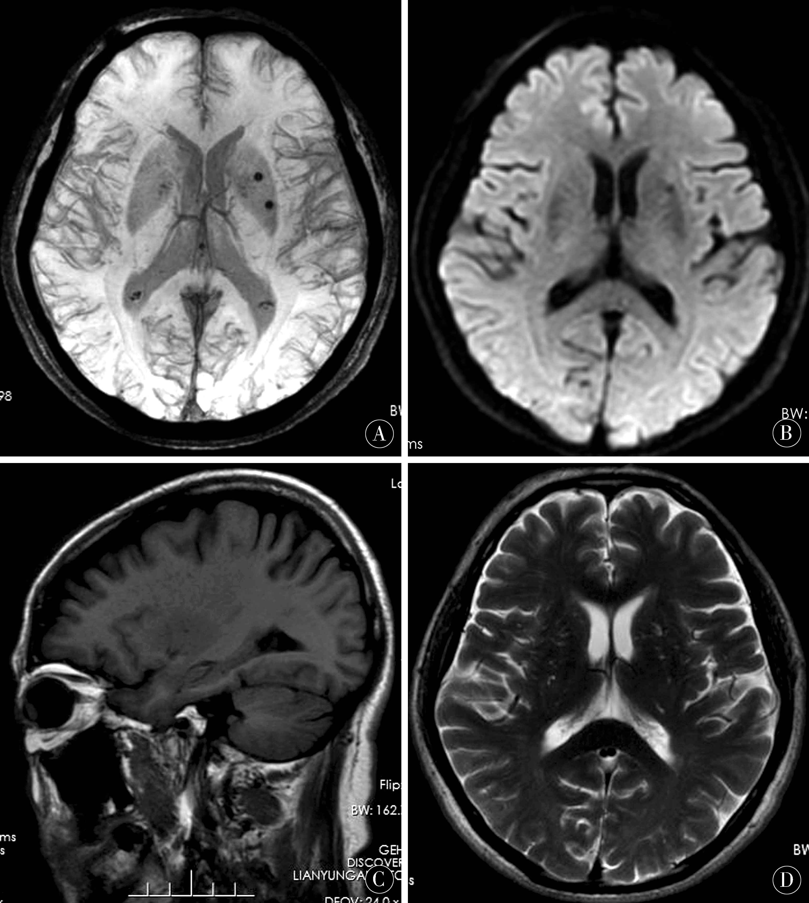
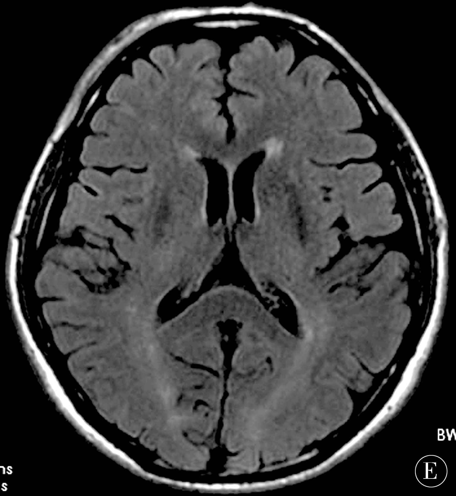
图1 女,65岁,缺血性脑卒中 A:SWI提示左侧基底节区两枚斑点状低信号,提示CMBs;B:DWI提示左侧基底节区一枚点状低信号;C~E:分别为T1WI、T2WI、T2FLAIR均未显示CMBs
Figure 1 Female,65 years old,with ischemic stroke A:SWI indicated two dotted hyposignals in left basal ganglia,indicating CMBs; B:DWI indicated one dotted hyposignal in left basal ganglia; C-E:T1WI,T2WI and T2FLAIR did not show CMBs,respectively
3 讨论
CMBs是由于脑微小血管发生纤维透明样变性等改变导致血管壁通透性增加,继而引发血细胞的渗出,导致含铁血红素于脑实质内沉积,其属于脑血管病范畴,其与高龄、高血压、脑出血、脑血管淀粉样变性等关系密切[8-12]。CMBs并不会引发神经细胞的坏死,但由于其引发胶质细胞的增殖和单核巨噬细胞的渗出,导致相应神经元功能障碍。CMBs是发生出血性脑卒中的独立性危险因素,与年龄成正比,且CMBs能够增加缺血性脑卒中患者溶栓后发生出血的风险,研究显示CMBs与各种类型的脑卒中均关系密切[13-15]。CMBs可用于预测出血性脑卒中发生的可能[16-18]。既往研究亦认为,皮层及皮层下CMBs是出血性脑卒中复发的独立危险因素。同时使用抗血小板药物可增加CMBs的发生,对脑血管病的二级预防具有指导意义[19-20],通过临床尽早采取针对性治疗能够减少其发生的概率。但CMBs患者多无典型临床表现,由于CMBs的体积较小,且病灶周围无明显水肿,导致临床常用影像检查方法中,包括CT及常规磁共振检查均不易显示,极易造成漏诊和误诊的发生[21],采取高效的影像检查方法则是患者诊断及治疗的前提基础。
磁共振检查中SWI序列是一种基于T2WI为基础,通过利用组织间磁化率差异进行成像的方法[22-23]。由于其对脱氧血红蛋白、正铁血红蛋白及含铁血黄素等顺磁性物资具有高度的敏感性,可同时获得磁距图和相位图。尽管CMBs病灶较小,但SWI对于其造成的局部磁场的不均匀仍能敏感检测,对于CMBs的检出明显高于常规检查方法[24-25]。同时也指出,SWI序列提示的点状或类圆形低信号反映的是顺磁性物质所产生的磁场,因此对于出血范围的显示会存在一定的夸大现象[26]。钙化和CMBs在SWI磁矩图均显示为低信号,需要结合相位图进行鉴别,两者在相位图上信号相反,钙化的反磁磁化率表现为低信号及以低信号为主的混合信号,CMBs的顺磁磁化率表现为高信号及以高信号为主的混合信号[27-28]。通过SWI可高效提高脑血管病患者CMBs病灶的检出率,为临床采取相关预防及治疗措施提供依据[29-30]。本研究显示,DWI、T1WI、T1WI及T2 FLAIR序列对于CMBs的检出均明显低于SWI序列,同时也发现DWI序列对与CMBs的检出高于其他常规序列,但仍低于SWI序列。同时本研究针对CMBs发生部位上亦发现CMBs分布与病因密切相关,基底节区及脑叶深部的CMBs多由高血压引起,多表现为类圆形或斑点状低信号,边缘光整,周围水肿不明显;缺血性脑卒中并发CMBs常表现为梗死区内或边缘点状、条片状低信号;皮层及皮层下CMBs多由脑淀粉样血管变性引发;弥漫性轴索损伤多表现为皮层下及基底节区多发斑点状低信号。
SWI序列能够高效显示CMBs,在其检出方面明显高于磁共振常规序列,具有明显的优势,同时不同病因导致的CMBs在形态及分布方面亦具有其一定的特征性表现。
4 参考文献
[1] AKOUDAD S,PORTEGIES M L,KOUDSTAAL P J,et al.Cerebral microbleeds are associated with an increased risk of stroke:the rotterdam study[J].Circulation,2015,132(6):509-516.
[2] FABOZZI F J,MCBBIDE J,CLANCY M.The Post-Crisis CMBS Market:Will Regulations Prevent Another Market Meltdown[J].J Portfolio Management,2015,41(6):118-125.
[3] LIU J,WANG D,XIONG Y,et al.A cohort study of relationship between serum calcium levels and cerebral microbleeds(CMBs) in ischemic stroke patients with AF and/or RHD[J].Medicine,2016,95(26):4 033.
[4] KLEINIG T J.Associations and implications of cerebral microbleeds[J].J Clin Neurosci,2013,20(7):919-927.
[5] HAACKE E M,LIU S,BUCH S,et al.Quantitative susceptibility mapping:current status and future directions[J].Magn Reson Imaging,2015,33(1):1-25.
[6] WANG S,DUAN C,ZHANG P,et al.Reaserch progress of quantitative susceptibility mapping in MRI[J].J Biomedical Eng,2015,32(5):1 131-1 134.
[7] SIRIN S,HUENING B,STENIN A,et al.improved detection of in tracranial hemorrhage in term and preterm neonates using SUS ceptibility weighted imaging[J].Arch Dis Child,2012,97(2):57-59.
[8] CHENG A L,BATOOL S,MCCREARY C R,et al.Susceptibility-weighted imaging is more reliable than T2*weighted gradientre called echo MRI for detecting microbleeds[J].Stroke,2013,44(10):2 782-2 786.
[9] HILAL S,MOK V,YOUN Y C,et al.Prevalence,risk factors and consequences of cerebral small vessel diseases:data from three Asian countries[J].J Neurol Neurosurg Psychiatry,2017,88(8):669-674.
[10] HUANG Y,MEI W L,LIU H Q,et al.Diagnostic value of clot burden score of susceptibility vessel sign in arterial thrombosis of acute ischemic stroke and its association with prognosis[J].Natl Med J China,2017,97(1):7-11.
[11] PAYAVASH S,BENSON J C,TALEB S,et al.Susceptible vessel sign:identification of arterial occlusion and clinical implications in acute ischaemic stroke[J].Clin Radiol,2017,72(2):116-122.
[12] KAPLAN E H,GOTTESMAN R F,LLINAS R H,et al.The association between specific substances of abuse and subcortical intracerebral hemorrhage versus ischemic lacunar infarction[J].Front Neurol,2014,5(5):174-176.
[13] TOTH P,TARANTINI S,ASHPOLE N M,et al.IGF-1 deficiency impairs neurovascular coupling in mice:implications for cerebromicrovascular aging[J].Aging Cell,20l5,14(6):1 034-1 044.
[14] AHN S J,ANRATHER J,NISHIMURA N,et al.Diverse inflammatory response after cerebral microbleeds includes coordinated microglial migration and proliferation[J].Stroke,2018,49(7):1 719-1 726.
[15] BATOOL S,PATIL S,NOSEWORTHY M D,et al.AbstractW MP119:sensitivity and reliahility of MRI SWI compared with GRE sequences for detecting microbleeds in a community population[J].Stroke,2015,11(4):13-17.
[16] RENARD D,WACONGNE A,LEFLOCH A,et al.Vicinity of FLAIR hyperintensities and SWI microbleeds in cerebral amyloid Angiopathy-Related inflammation[J].Eur Neurol,2016,75(5/6):223-224.
[17] KOO D L,KIM J Y,LIM J S,et al.Cerebral Microbleeds on MRI in Patients with Obstructive Sleep Apnea[J].J Clin Sleep Med,2017,13(1):65-72.
[18] PARK M G,OH S J,BAIK S K,et al.Susceptibility-weighted Imaging for Detection of Thrombus in Acute Cardioembolic Stroke[J].Stroke,2016,18(1):73-97.
[19] UNGVARI Z,TARANTINI S,KIRKPATRICK A C,et al.Cerebral microhemorrhages:mechanisms,consequences,and prevention[J].Am J Physiol Heatr Circ Physiol,2017,312(6):128-143.
[20] LIU C,LI W,TONG K A,et al.Susceptibility-weighted imaging and quantitative susceptibility mapping in the brain[J].J Magn Reson Imaging,2015,42(1):23-41.
[21] ROMERO J R,BEISER A,HIMALI J J,et al.Cerebral microbleeds and risk of incident dementia:the Framingham Heart Study[J].Neurobiol Aging,2017,54(7):94-99.
[22] ZHAO G,SUN L,WANG Z,et al.Evaluation of the role of susceptibility weighted imaging in thrombolytic therapy for acute ischemic stroke[J].J Clin Neurosci,2017,40(1):175-179.
[23] HSU C C,KWON G N C,HAPUGODA S,et al.Susceptibility weighted imaging in acute cerebral ischemia:review of emerging technical concepts and clinical applications[J].Neuroradiol J,2017,30(2):109-119.
[24] BARBOSA J H,SANTOS A C,SALMON C E.Susceptibility weighted imaging:Differentiating between calcification and hemosiderin[J].Radiol Bras,2015,48(2):93-100.
[25] MALKKI H.Cerebrovascular disorders:Cerebral microbleeds linked to increased risk of cognitive decline[J].Nat Rev Neurol,2016,12(4):432.
[26] TSIVGOULIS G,ZAND R,KATSANOS A H,et al.Risk of symptomatic intracerebral hemorrhage after intravenous thrombolysis in patients with acute ischemic stroke and high cerebral microbleed burden;a Meta-analysis[J].JAMA Neurol,2016,73(6):675-683.
[27] BOURCIER R,VOLPI S,GUYOMARCH B,et al.Susceptibility vessel sign on MRI predicts favorable clinical outcome in patients with anterior circulation acute stroke treated with mechanical thrombectomy[J].AJNR AM Neuroradiol,2015,36(12):2 346-2 353.
[28] LOU Y,GONG Z,ZHOU Y,et al.Increased susceptibility of asymmetrically prominent cortical veins correlates with misery perfusion in patients with occlusion of the middle cerebral artery[J].Eur Radiol,2017,27(6):2 381-2 390.
[29] BERGERON S,CHEN Y,AUGER F,et al.Role of cortical microbleeds in cognitive impairment:in vivo behavioral and imaging characterization of a novel murin model[J].J Cereb Blood Flow Metal,2018,24(9):46-49.
[30] 陶永君.磁敏感加权成像在脑微出血诊断中的应用价值[J].中国实用神经疾病杂志,2015,18(8):96-97.
(收稿2019-01-02)
本文责编:关慧
本文引用信息:李士坤,符大勇,卢明聪,周建国,马先军,刘晓丽.磁敏感成像技术在脑微出血诊断中的价值研究[J].中国实用神经疾病杂志,2019,22(1):46-50.DOI:10.12083/SYSJ.2019.01.009
Reference information:LI Shikun,FU Dayong,LU Mingcong,ZHOU Jianguo,MA Xianjun,LIU Xiaoli.Application value of magnetic sensitive imaging in the diagnosis of cerebral microhemorrhage[J].Chinese Journal of Practical Nervous Diseases,2019,22(1):46-50.DOI:10.12083/SYSJ.2019.01.009